| Data Images |
 |
|
|
|
|
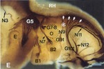
| Fig 6E Mark et al, 1993 [PMID:8287791] .
Copyright: This image is from Development and is displayed with the permission of the Company of Biologists Ltd who owns the copyright. |
|
|
|
|
|
|
| Expression pattern clarity: |
 |
| Find spatially similar wholemount expression patterns: |
 |
|
|
| Notes: |
|
Image annotations: B1 and B2, 1st and 2nd pharyngeal (branchial) arches; E, eye; G5, ganglion of cranial nerve V (Gasser's ganglion); G7-8, facial-acoustic ganglion; G9t-G10t, inferior (trunk) ganglia of cranial nerves IX and X respectively; ; N3, N7, N9, N11, N12 are cranial nerves III, VII, IX, XI and XII respectively; O, otocyst; RH, rhombencephalon; asterisk, rostral level of the superior ganglionic complex of the cranial nerves IX and X; small white arrows, roots of cranial nerves IX and X. The dorsal portion of the otocyst was removed to unmask the pial surface of the rhombencephalon. The position of the otocyst is indicated by a dashed line. |
|
| Expression Pattern Description |
| Spatial Annotation: |
 | | | | | Annotation colour key:
 |
strong |
 |
moderate |
 |
weak |
 |
possible |
 |
not detected |
|
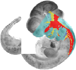
| wholemount mapping | | | | |
| Download individual expression domains: |
|
| Download all expression domains: |
EMAGE:1244_all_domains.zip |
| Find spatially similar wholemount expression patterns: |
 |
| Morphological match to the template: |
 |
|
| Text Annotation: |
| Structure | Level | Pattern | Notes |
|---|
| embryo |
 |
detected |
| regional | Antibody 2H3 was used to visualize neurons |
|
| Annotation Validation: |
EMAGE Editor |
|
| Detection Reagent |
| Type: | antibody |
| Identifier: | MGI:1330766 |
| Entity Detected: | Nefm, neurofilament, medium polypeptide ( MGI:97314) |
| Notes: | The anti-NEF3 antibody used in this study by Mark et al, 1993 [PMID:8287791] is described as "2H3 (a mouse monoclonal antibody against the 155000 Mr neurofilament protein; Developmental Studies Hybridoma Bank, Departmental Pharmacology of Molecular Sciences, John Hopkins University School of Medicine, Baltimore, MD, USA)". Details below are taken from the Developmental Studies Hybridoma Bank online catalogue. |
| Antibody Type: | monoclonal |
| Raised In: | mouse |
| Supplier: | Developmental Studies Hybridoma Bank |
| Catalogue Number: | 2H3 |
|
| Specimen |
| Organism: | mouse |
| Age: | 11.0 dpc |
| Theiler Stage: | TS18 |
| Mutations: | none (wild-type) |
| Preparation: | wholemount |
|
| Procedures |
| Fixation: | 3% paraformaldehyde |
| Secondary Antibody: | peroxidase-conjugated goat anti-mouse IgG |
| Labelled with: | horse radish peroxidase |
| Visualisation method: | 4-chloro-1 naphtol (Merck, Darmstadt, FRG) |
|
| General Information |
| Authors: | Mark et al, 1993 [PMID:8287791]
Indexed by GXD, Spatially mapped by EMAGE |
| Submitted by: | EMAGE EDITOR, Institute of Genetics and Molecular Medicine, Western General Hospital, Crewe Road, Edinburgh, UK EH4 2XU |
| Experiment type: | non-screen |
| References: | [ PMID:8287791] Mark M, Lufkin T, Vonesch JL, Ruberte E, Olivo JC, Dolle P, Gorry P, Lumsden A, Chambon P 1993 Two rhombomeres are altered in Hoxa-1 mutant mice. Development (119):319-38 |
| Links: | MGI:1343248 same experiment |
| | Ensembl same gene |
| | Allen Brain Atlas same gene |
| | BioGPS same gene |
| | International Mouse Knockout Project Status same gene |
| | GEISHA Chicken ISH Database same gene |
| | EMBL-EBI Gene Expression Atlas same gene |
| | BrainStars same gene |
| | ViBrism same gene |
| Data Source |  |
|
 icon to keep this page displayed.)
 icon to keep this page displayed.)