
Theiler Stage Definition
| Theiler Stage | Summary | Anatomy Ontology | Theiler Book Text (pdf) | dpc (range)1 | Somite Number2 | Cell Number | (C57BLxCBA) F1 mice3 | PO Mice |
|---|---|---|---|---|---|---|---|---|
| 1 | 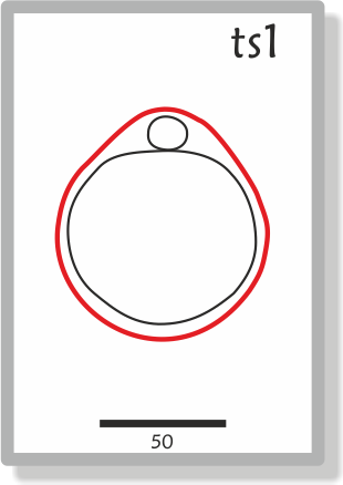 |
TS01 Anatomy | TS01 Theiler Description | 0-0.9 (0-2.5) | 1 | One-cell egg | ||
| 2 | 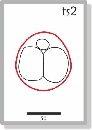 |
TS02 Anatomy | TS02 Theiler Description | 1 (1-2.5) | 2-4 | Dividing egg | ||
| 3 | 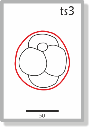 |
TS03 Anatomy | TS03 Theiler Description | 2 (1-3.5) | 4-16 | Morula | ||
| 4 | 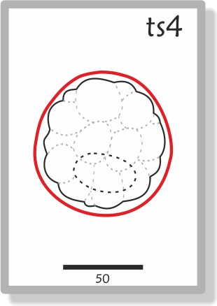 |
TS04 Anatomy | TS04 Theiler Description | 3 (2-4) | 16-40 | Blastocyst, Inner cell mass apparent | ||
| 5 | 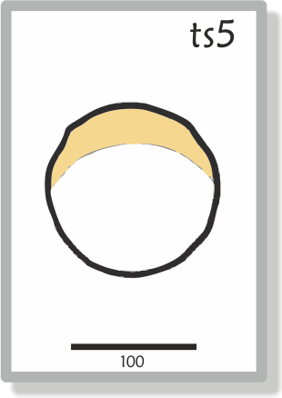 |
TS05 Anatomy | TS05 Theiler Description | 4 (3-5.5) | Blastocyst (zona free) | |||
| 6 | 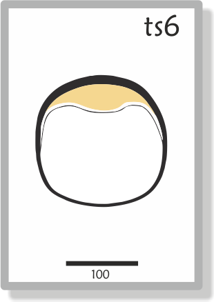 |
TS06 Anatomy | TS06 Theiler Description | 4.5 (4-5.5) | Attachment of blastocyst, primary endoderm covers blastocoelic surface of inner cell mass | |||
| 7 | 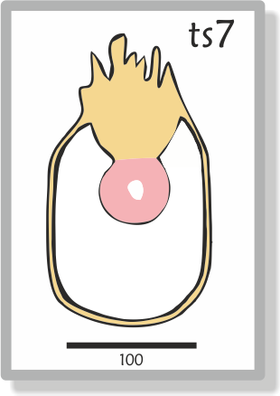 |
TS07 Anatomy | TS07 Theiler Description | 5 (4.5-6) | Implantation and formation of egg cylinder Ectoplacental cone appears, enlarged epiblast, primary endoderm lines mural trophectoderm | |||
| 8 | 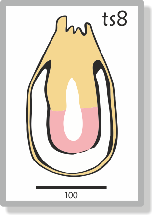 |
TS08 Anatomy | TS08 Theiler Description | 6 (5-6.5) | Differentiation of egg cylinder Implantation sites 2x3mm. Ectoplacental cone region invaded by maternal blood, Reichert's membrane and proamniotic cavity form | |||
| 9a | 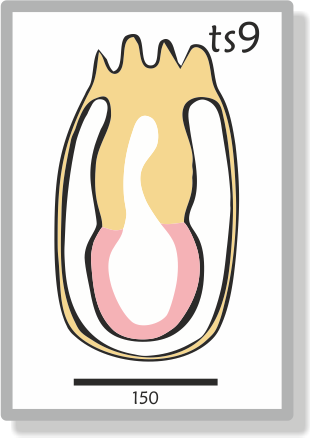 |
TS09 Anatomy | TS09 Theiler Description | 6.5 (6.25-7.25) | Pre-streak(PS), advanced endometrial reaction, ectoplacental cone invaded by blood, extraembryonic ectoderm, embryonic axis visible | PS | ||
| 9b | 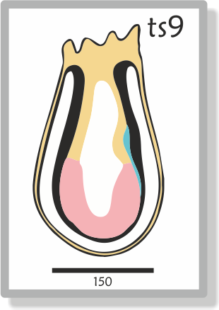 |
Early streak(ES), gastrulation starts, first evidence of mesoderm | ES | |||||
| 10a | 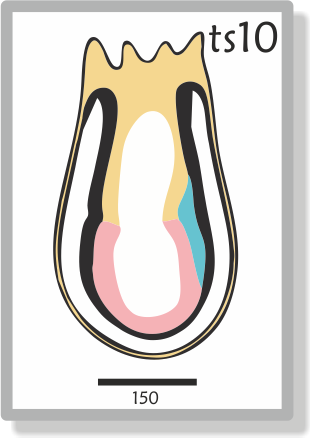 |
TS10 Anatomy | TS10 Theiler Description | 7 (6.5-7.75) | Mid streak (MS), amniotic fold starts to form | MS | ||
| 10b | 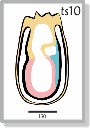 |
Late streak, no bud (LSOB), exocoelom | LS | |||||
| 10c | 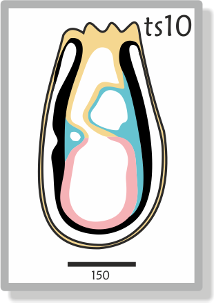 |
Late streak, early bud (LSEB), allantoic bud first appears, node, amnion closing | ||||||
| 11a | 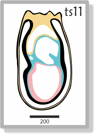 |
TS11 Anatomy | TS11 Theiler Description | 7.5 (7.25-8) | Neural plate (NP), head process developing, amnion complete | OB | ||
| 11b | 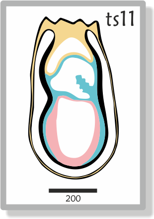 |
Late neural plate (LNP), elongated allantoic bud | EB/LB | |||||
| 11c | 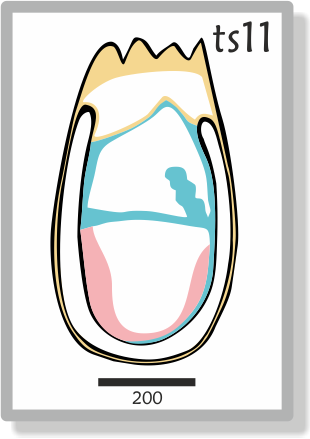 |
Early head fold (EHF) | EHF | |||||
| 11d |  |
Late head fold (LHF), foregut invagination | LHF | |||||
| 12a |  |
TS12 Anatomy | TS12 Theiler Description | 8 (7.5-8.75) | 1-4 | 1-4 somites, allantois extends, 1st branchial arch, heart starts to form, foregut pocket visible, preotic sulcus at 2-3 somite stage) | ||
| 12b | 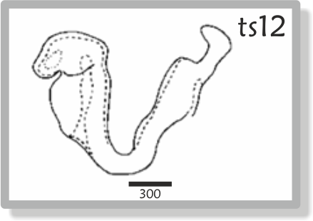 |
5-7 | 5-7 somites, allantois contacts chorion at the
end of TS12 Absent 2nd arch, >7 somites |
|||||
| 13 | 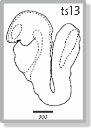 |
TS13 Anatomy | TS13 Theiler Description | 8.5 (8-9.25) | 8-12 | Turning of the embryo, 1st branchial arch has
maxillary and mandibular components, 2nd arch present Absent 3rd branchial arch |
||
| 14 |  |
TS14 Anatomy | TS14 Theiler Description | 9 (8.5-9.75) | 13-20 | Formation & closure of ant. neuropore, otic
pit indented but not closed, 3rd branchial arch visible Absent forelimb bud |
||
| 15 |  |
TS15 Anatomy | TS15 Theiler Description | 9.5 (9-10.5) | 21-29 | Formation of post. neuropore, forelimb bud, forebrain
vesicle subdivides
Absent hindlimb bud, Rathke's pouch |
||
| 16 | 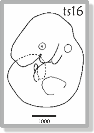 |
TS16 Anatomy | TS16 Theiler Description | 10 (9.5-10.75) | 30-34 | Posterior neuropore closes, Formation of hindlimb
& tail buds, lens plate, Rathke's pouch; the indented nasal processes
start to form
Absent thin & long tail |
||
| 17 | 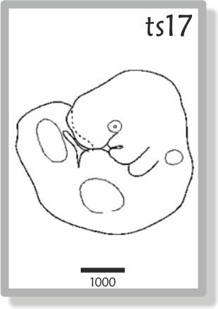 |
TS17 Anatomy | TS17 Theiler Description | 10.5 (10-11.25) | 35-39 | Deep lens indentation, adv. devel. of brain tube,
tail elongates and thins, umbilical hernia starts to form
Absent nasal pits |
||
| 18 | 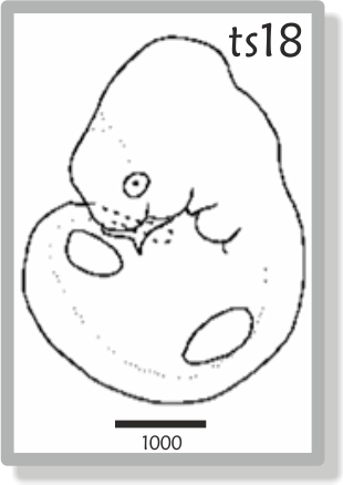 |
TS18 Anatomy | TS18 Theiler Description | 11 (10.5-11.25) | 40-44 | Closure of lens vesicle, nasal pits, cervical
somites no longer visible
Absent auditory hillocks, anterior footplate |
||
| 19 | 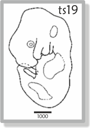 |
TS19 Anatomy | TS19 Theiler Description | 11.5 (11-12.25) | 45-47 | Lens vesicle completely separated from the surface epithelium, Anterior, but no posterior, footplate. Auditory hillocks first visible
Absent retinal pigmentation and sign of fingers |
||
| 20 | 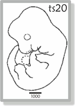 |
TS20 Anatomy | TS20 Theiler Description | 12 (11.5-13) | 48-51 | Earliest sign of fingers, (splayed-out), posterior
footplate apparent, retina pigmentation apparent, tongue well-defined, brain vesicles clear
Absent 5 rows of whiskers, indented |
||
| 21 | 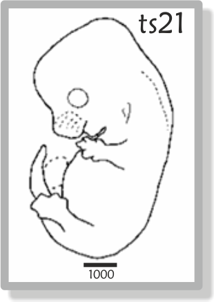 |
TS21 Anatomy | TS21 Theiler Description | 13 (12.5-14) | 52-55 | Anterior footplate indented, elbow and wrist
identifiable, 5 rows of whiskers, umbilical hernia now clearly apparent
Absent hair follicles, fingers separate distally |
||
| 22 | 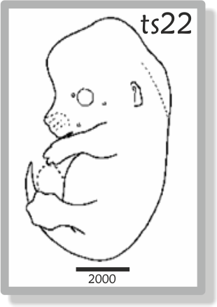 |
TS22 Anatomy | TS22 Theiler Description | 14 (13.5-15) | 56-~60 | Fingers separate distally, only indentations
between digits of the posterior footplate, long bones of limbs present,
hair follicles in pectoral, pelvic and trunk regions
Absent open eyelids, hair follicles in cephalic region |
||
| 23 |  |
TS23 Anatomy | TS23 Theiler Description | 15 | Fingers & Toes separate, hair follicles also
in cephalic region but not at periphery of vibrissae, eyelids open
Absent nail primordia, fingers 2-5 parallel |
|||
| 24 | 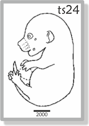 |
TS24 Anatomy | TS24 Theiler Description | 16 | Reposition of umbilical hernia, eyelids closing,
fingers 2-5 are parallel, nail primordia visible on toes
Absent wrinkled skin, fingers & toes joined together |
|||
| 25 | 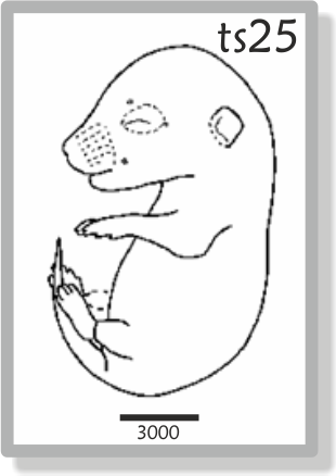 |
TS25 Anatomy | TS25 Theiler Description | 17 | Skin is wrinkled, eyelids are closed, umbilical hernia is gone
Absent ear extending over auditory meatus, long whiskers |
|||
| 26 | 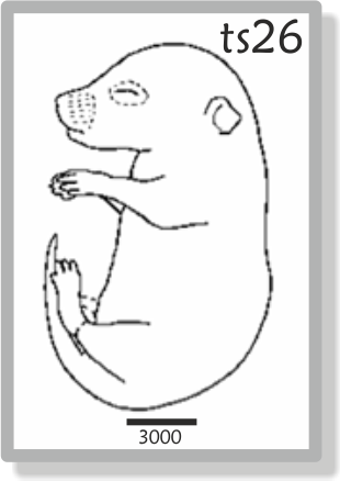 |
TS26 Anatomy | TS26 Theiler Description | 18 | Long whiskers, eyes barely visible through closed eyelids, ear covers auditory meatus | |||
| 27 | 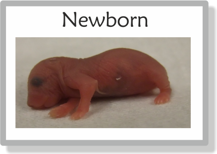 |
TS27 Theiler Description | 19 | Newborn Mouse | ||||
| 28 | 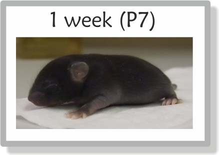 |
TS28 Theiler Description | Postnatal development | |||||
| Adult | 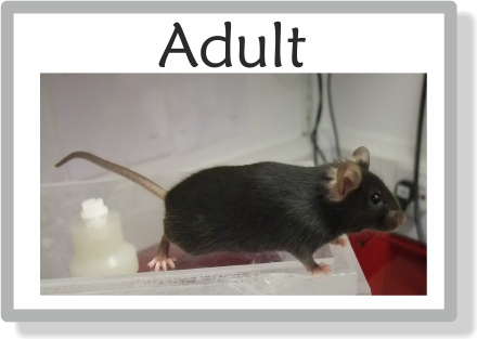 |
MGI anatomical dictionary | Adult Mouse |
Comments
Mouse embryos can be staged according to a variety of criteria, the most general of which are those described by Theiler in "The House Mouse: Atlas of Mouse Development" (Springer-Verlag, New York, 1989). Theiler's criteria are too broad to distinguish many of the important phases of early development and must therefore be supplemented by others, for example, cell number, somite number or those charcteristics used by Downs and Davies (1993) Development, 118, 1255. We have therefore combined these different criteria in the table which defines a new set of stages based on the numbered Theiler series, but with intermediate divisions indicated by non-integer stage numbers5. Embryos of the same gestational age may differ in their stage of development. We have therefore included in the table an indication of the expected range of gestational ages (days of gestation, dpc) over which each developmental stage may be found. Different mouse strains develop at different rates and, in some cases show differences in the relative rates of development of different organs. Strictly, the stages recognised by Downs and Davies apply to outbred mice of the PO strain. The data in the remainder of the table below refer to embryos of crosses between F1 hybrid (C57BL X CBA) mice.
General comment on timing (dpc): In judging the lower and upper ranges of dpc equivalent to a particular Theiler stage, we have generally followed Theiler's book and, in most cases, have given a wider range than Theiler, because the numbers of embryos he cites are small. We have given a larger range at the maximum than the minimum because, in general, embryos are more likely to be retarded by their environment or genetic constitution than made to proceed more quickly through development. In most cases, however, the resulting dpc range is an estimate that is consistent with the results of Theiler, but not based on additional evidence.
Comment on somite numbers: The range of somite numbers for each stage is given only as a guide to what might be expected of typical embryos. As can be seen from Theiler (1989)3 the true range can be much wider. Therefore, for all stages after TS12, the somite number should not be taken as a reliable global indicator of the overall embryo stage.
1.dpc(range) Days post conception, with the morning after the vaginal plug is found being designated 0.5 dpc (or E0.5). For detailed discussion see Kaufman (1994). The Atlas of Mouse Development (2nd printing), pp. 515-525. London: Academic Press.
2. Somite Number The figure given refers to the number of the most caudal somite. No account is taken of somites partitioning into dermomyotomes and sclerotomes, nor of their subsequent differentiation.
3.(C57BLxCBA) F1 mice Adapted from Theiler (1989) [The House Mouse: Atlas of Embryonic Development. New York: Springer-Verlag] and Kaufman (1994); detailed staging for Theiler stages 9-12 courtesy of K. Lawson [personal communication].
4. PO Mice From Downes, K.M. and Davies, T. (1993). Staging of gastrulating mouse embryos by morphological landmarks in the dissecting microscope. Development, 118, 1255 - 1266.
5. Reference Bard, J.B.L., Kaufman, M.H., Dubreuil, C., Brune. R.M., Burger, A., Baldock, R.A., Davidson, D.R. (1998). An internet-accessible database of mouse developmental anatomy based on a systematic nomenclature. Mechanisms of Development 74, 111-20.
Richard Baldock, Jonathan Bard, Duncan Davidson and Kirstie Lawson, 7th May 1998


