
"Morphogenesis": exploring the 3D animation possibilities using data obtained with the imaging technique Optical Projection Tomography (OPT).
Dr Chris Armit, Glasgow School of Art.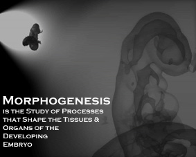
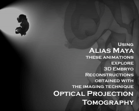
Morphogenesis is a collaborative effort between the Mouse Atlas Project at the MRC Human Genetics Unit and the Glasgow School of Art that aims to explore the 3D animation possibilities that are achievable using data obtained with the imaging technique Optical Projection Tomography (OPT).
The aims of Morphogenesis were threefold:
- To discern to what degree 3D volumetric data sets (OPT data) could be incorporated into 3D animations
- To use the data as the basis of an informative introduction for a short web-based documentary on the subject of embryonic development
- To use the data as the basis of an abstract animation
Volumes and Surfaces
For manipulation of OPT data in commercial 3D animation
packages it is necessary to generate surfaces from the volume data.
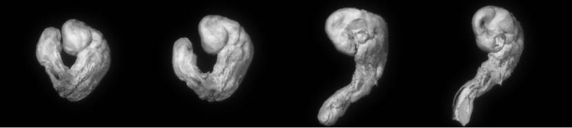
The surfaces generated are difficult to animate as a character in commercial 3D animation packages such as Alias Maya. The reason for this is that they are not in a conventional ‘bind pose’ with the tail unravelled and pointing straight downwards.
One method to overcome this problem is to preprocess the entire volume data set such that it is unravelled to suit the animator’s need*. Below is this principle applied to one of the OPT data sets.
Click on links to see movies
| Movie 1 | AVI (1.32MB) | MPEG (2.51MB) |
| Movie 2 | AVI (1.32MB) | MPEG (2.61MB) |
| Movie 3 | AVI (1.32MB) | MPEG (2.61MB) |
Genes and Embryos
is a short web-based documentary on the subject of embryo development. It is aimed at the informed layman who may wish to know more about the relationship between genes and animal form.
For ease in downloading, this feature is divided into 3 movie files.
| Introduction | AVI (14.3MB) | MPEG (26.8MB) |
| Existence of Morphogens | AVI (22.MB) | MPEG (33.7MB) |
| Evolution of the Limbs | AVI (22.2MB) | MPEG (36.2MB) |
Note:- movie files contain both sound and images. I am very grateful to David bernard who allowed me to use his music in this production.
 |
The study of genes advances our understanding of how we develop from a single cell into a thoroughly recognisable human being. Many genes are switched on - or expressed during embryogenesis and these genes may ultimately serve to pattern the embryo. In this video clip,Professor Nick Hastie introduces this topic of genes and development whilst Dr Bob Hill discusses the impact of mutation to our understanding. |
 |
The concept of the morphogen is an important onein Developmental Biology. That a diffusible substance - a signal - may pattern surrounding tissue has its roots in regeneration experiments involving Hydra; from over 100 years ago. In the past 10 years, there has been a lot of interest in a protein - that encoded by the Sonic Hedgehoggene - which has many hallmarks of a morphogen. In particular, there is interest as to whether it is responsible for producing the correct sequence of finger or toes in the developing limb. In this video clip, Professor Lewis Wolpert and Dr Bob Hill discuss the existence of morphogens and address the possibility of Sonic Hedgehog actually being the limb morphogen. |
 |
It is believed that the limbs of land-based tetrapods (vertebrates with four feet) evolved from the fins of fish. How this transition took place is unclear, but may be related to changing patterns of gene expression. In this video clip, Dr Bob Hill discusses the evolution of the limb and how an understanding of Evolutionary and Developmental Genetics (EvoDevo) is increasing our understanding of how evolutionary change is achieved. |
Abstract Animations
Click on the links to download the movie files.
AVI (10.8MB) MPEG (27.2MB)
If one considers the artwork of Hieronymus Bosch or of Salvador Dali, of Remedios Varo or of Kay Sage, one may note that the image of either the egg or the embryo is often used symbolically in art. Despite the dynamism that may accompany these images, the use of the embryo as a symbolic archetype overlooks the observed feature that the embryo is itself a dynamic form, a shapeshifter whose contours change with advancing development. Each image of an embryo is itself a snapshot in time, and with advancing development the form of the embryo itself gradually becomes the form of the infant. Likewise, early stages of embryogenesis are not necessarily identifiable as embryos, their forms being abstract and in some instances representational of other animal forms.
It is widely believed that the forms of the developing embryo have something to do with genes. The language of development is that of measured doses of proliferation, death, and migration of the constituent cells, and the behaviour of these cells is itself dictated by the sets of genes which are switched on, or expressed therein. This new anatomy of gene expression often precedes the formation of tissue proper and can be seen as a series of templates over which tissue organises itself. In this conceptual model, the complexity of development relates to the sheer number of genes involved, which is estimated at somewhere between 30,000-40,000 genes in a mammal such as ourselves.
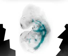
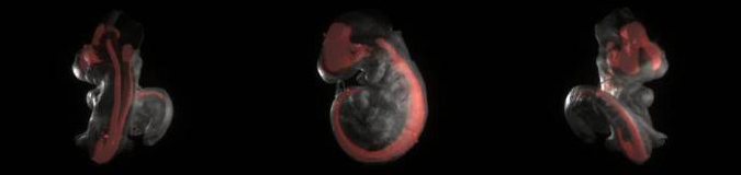
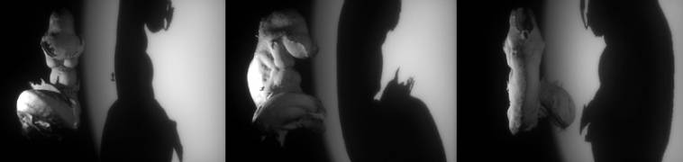
In the accompanying abstract animation, the author wishes to explore the form of the mid-stage mouse embryo, incorporating specifically those stages at which it is most similar to the human embryo. Representations of this ambiguous creature are created both in utero and in isolation, and surface renders of gene expression patterns are included where appropriate. In addition, the author wishes to recreate in a succession of scenes the OPT scanning procedure, the method by which the 3D reconstructions are generated. It is the author’s intention that this succession of shots will visually communicate some of the methods employed by researchers in the field.
For further information contact Chris Armit


