
Investigating effects of Pax6 on cell mixing in SeyNeu/Sey<-> +/+ chimaeric fetal mouse eyes using 3-dimensional reconstruction techniques.
Authors : Jane C. Quinn, Margaret H. Stark, Duncan Davidson, and Richard A. Baldock.
Reference : Quinn JC, West JD and Hill RE. Multiple functions for Pax6 in mouse eye and nasal development. Genes and Development V10 (1996) pp435-446 (PubMed)
Pax6 is a important gene involved in development of the eye, brain and nasal tissues. Mutations within the Pax6 gene in both mouse and man result in eye abnormalities. In the mouse, Pax6 lesions are responsible for the semi-dominant Small eye phenotype. To investigate roles for Pax6 in eye development, foetal mouse chimaeras were produced by aggregation of embryos compound heterozygous for mutations within the Pax6 gene (SeyNeu/Sey) to embryos wild-type (+/+) at the Pax6 locus. To allow positional visulaisation of SeyNeu/Sey cells unable to produce a functional Pax6 product, embryos derived from Small eye parents (SeyNeu/+ x Sey/+) contained a single copy of a reiterated beta-globin transgene were used for aggregation to +/+ embryos. Chimaeric foetuses were examined at E12.5. Wild-type and SeyNeu/Sey cell populations were differentiated within foetal tissues by positive identification of SeyNeu/Sey cells by DNA-DNA in-situ hybridisation to the beta-globin transgene.
Analysis of SeyNeu/Sey<-> +/+ chimaeric fetuses showed that SeyNeu/Sey cells were excluded from both the lens and nasal epithelium. Also, both of these tissues were always smaller and sometimes absent in SeyNeu/Sey<-> +/+ chimaeras containing a high proportion of mutant cells. Morphology of the optic cup was also severely affected, SeyNeu/Sey cells were excluded from the retinal pigment epithelium and did not intermix with +/+ cells in other regions of the optic cup (Quinn, West and Hill, 1996).
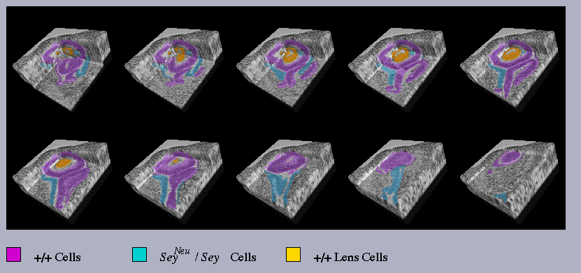
| Figure 1. a) | b) |
 |
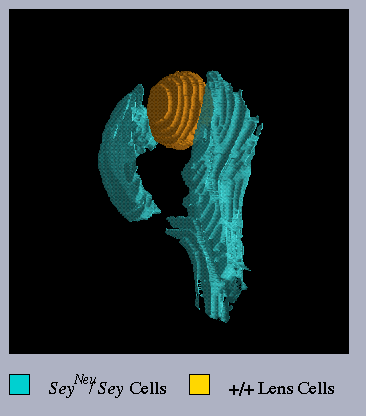 |
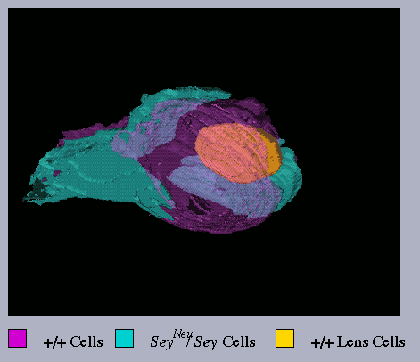
Figure 1. a) 3-D reconstruction of left eye from
SeyNeu/Sey<-> +/+
chimaera JC48 showing areas of +/+ cells - pink, SeyNeu/Sey
cells - blue and wild-type cells forming the lens- yellow. SeyNeu/Sey
and +/+ cells are observed to segregate into highly distinct but
confluent tissue areas constituting a relatively normal optic cup structure
containing a small lens. b) The same reconstruction as shown in
a) with areas of +/+ cells removed to show extent of SeyNeu/Sey
cell contribution to the developing optic cup.
| Figure 2. a) | b) |
 |
 |
Figure 2. 3-D 'excavation' reconstructions of a) left
and b) right eyes of SeyNeu/Sey<->
+/+ chimaera JC48. This technique allows viewing of the grey-level
image of the original histological sections together with the painted domains
by removal of 28mm sections from within the
whole 3-D object. This allows visualisation of the painted domains both
within context of surrounding tissues and in internal and external relationship
to each other.
Figure 2. a) Left eye of SeyNeu/Sey<->
+/+ chimaera JC48 showing that the abnormal eye structure has maintained
contact with the overlying surface ectoderm, note also the lens appears
partially enclosed within the +/+ optic cup tissue. b) The right
eye of SeyNeu/Sey<-> +/+
chimaera JC48 showing highly convoluted optic cup morphology with loss
of lens formation, this abnormal structure is also maintaining contact
with the overlying surface ectoderm.
By using the 3-D excavation technique for visualisation of these chimaeric
eye structures it was possible to compare morphology between the left and
right eyes of SeyNeu/Sey<->
+/+ chimaera. Examination of the left eye of chimaera JC48 (Figure
2. a) showed that where lens formation had occurred, +/+ and SeyNeu/Sey
cells were able to contribute to both proximal and distal regions of
the developing optic cup although SeyNeu/Sey
cells were mainly confined to the outer layer. In comparison, loss
of lens formation (Figure 2. b) resulted in a highly disorganised optic
cup morphology where SeyNeu/Sey cells
appeared to be restricted to the more proximal (stalk) regions of the optic
cup.
In conclusion, using the 3-D reconstruction techniques it was possible to see that areas of +/+ or SeyNeu/Sey optic cup tissue were considerably larger than had been intimated from histological sections alone. These segregated optic cup tissue areas were found to be confluent even within highly abnormal optic cup structures. In addition, SeyNeu/Sey cells were observed to be mainly restricted to the outer layer and more proximal optic cup regions, this effect being marked in the absence of lens development. Therefore, loss of a functional Pax6 product disrupts cell-to-cell interactions and perturbs the ability of SeyNeu/Sey cells to form distal optic cup strutures in the eyes of SeyNeu/Sey<-> +/+ chimaeras.


