

The Edinburgh Mouse Atlas Project: School Tutorial - Pictures and Movies
Click on the picture to get a bigger version or to see the movie sequence.Pictures
 |
Selected anatomical domains of the 9-day embryo model (Stage 14) displayed in section mode with over-laid anatomical components. Colours are re-used for different painted domains, these images show the resolution of the Mouse Atlas painted domains. |
 |
Stereo-pair image showing selected vessels of the 9-day embryo model. The stereo view can be seen by crossing your eyes and merging the two images, either with this view or the enlarged version. The angular separation between the two views is 12 degrees. |
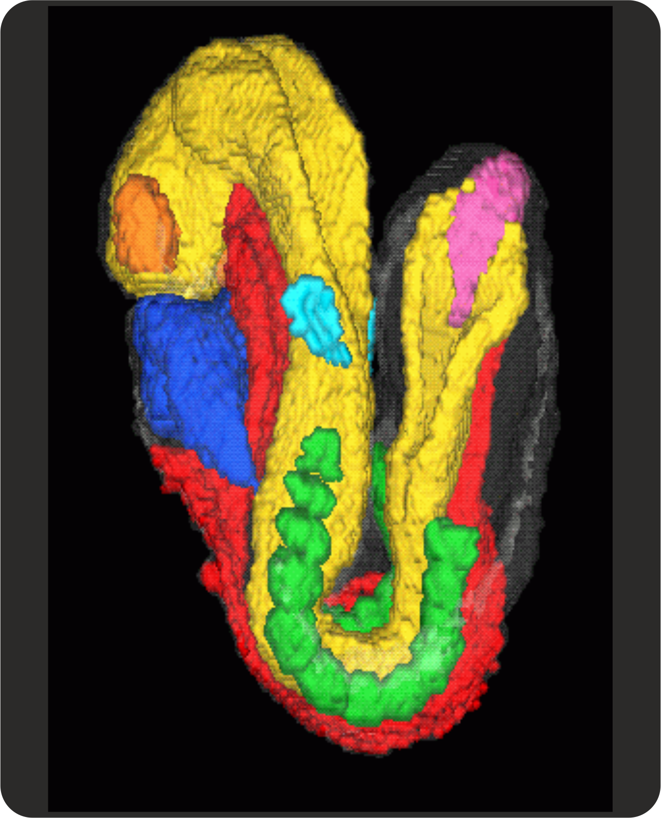 |
This pseudo-3D view of the 8.5-day old embryo (Stage 13) displays the painted domains of the neural tube (yellow), gut (red) heart (blue) somites (green) ear placode (pale blue) and streak (mauve). These tissues of the embryo are enclosed in translucent surface ectoderm. |
Movies
This movie shows an animation of all the grey-level sections from which the 3-D reconstruction of the 9-day embryo model (Stage 14) was made. |
|
Rotating view of selected 9-day anatomy with a grey-level section |
|
Striptease - mouse anatomy revealed - from the skin inwards though! |
|
The vascular system |
|
Neural and other tissues |
|
A volume rendered view of the mouse - a bit like an X-ray. |


