

The Edinburgh Mouse Atlas Project: School Tutorial - Sectioning the Mouse Embryo
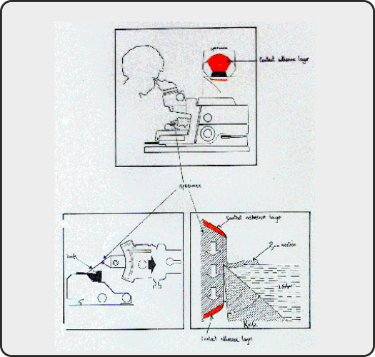 |
The mouse embryos are very small in the early stages and biologists need to locate their data to within about 10 microns. This means that we need a reconstruction that has come from serial sections - very thin, consecutive slices of the embryo that can be stained to make them visible under the microscope. This is done by embedding the embryo in wax or plastic resin (Araldite) then cutting the block using a microtome |
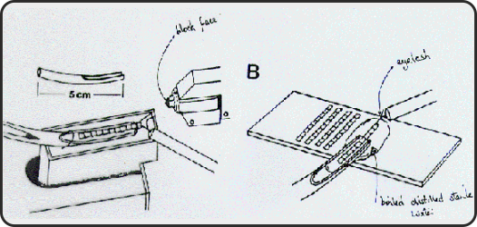 |
Getting a full set of sections is sometimes really difficult and at the Human Genetics Unit a highly specialized technique is used. As the string of sections is cut it is floated in a channel of water to keep it straight. (The string of sections is so fine that it has to be manipulated using an eyelash taped to a cocktail stick!) |
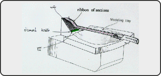 |
The ribbon of sections is then put onto slides where they are stained and can be viewed under the microscope. |


