mMRI
mMRI Microscopic magnetic resonance imaging is probably the most technically-complex of the techniques I have listed here. I will therefore not attempt to explain how it works, as there are other web-sites dedicated to MRI (for example the BioImaging Center at Caltech). The important aspect with respect to other imaging techniques, is that it requires placing the specimen inside a strong magnetic field. MRI is becoming increasingly common as a medical imaging technique for large specimen (brain scans etc.), however as the intended imaging scale gets smaller, the strength of the magnetic field needed becomes greater. To image specimen a few millimeters across requires a magnet which is very strong and therefore very expensive.
The advantages which mMRI displays are that it can image specimen which are too large and opaque for OPT microscopy, and that it can image living specimen.
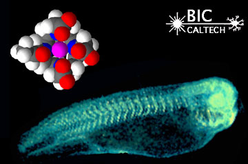 Section through a living tadpole, in which expression of the LacZ reporter gene is detected using the high-contrast agent whose molecular structure is shown above. This molecule is a substrate for the b-galactosidase enzyme (expressed from the LacZ reporter gene) which displays high MRI contrast after cleavage of its galactose side-chain.
However OPT microscopy displays the following advantages over mMRI:
1. It can image coloured stains. This allows us to image data from:
- Normal in-situ hybridisation experiments (using the BCIP and NBT substrates)
- Reporter gene experiments (such as LacZ and alkaline phosphatase)
2. It can image fluorescent dyes. This allows us to image:
- Multiple signals from the same tissue
- Standard fluorescent antibody reagents
3. Only takes about 15 minutes to image each channel. This speed allows us to image many specimen rapidly, and therefore to perform studies of natural variation in shape, gene expression, mutant phenotypes etc.
4. It is much cheaper than MRI, and works in conjunction with a normal "dissecting" microscope, making it accessible to many more biologists.
5. It images at a higher resolution than MRI (see following figures).
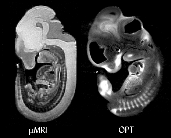 Comparison between mMRI and OPT. In both cases the specimen is an E11.5 mouse embryo. The mMRI image shows higher contrast between tissues, while OPT shows greater resolution. The OPT-scanned embryo was also stained for the expression of Sox9 with a coloured dye (BCIP/NBT), which can be seen as the white regions. mMRI cannot image common assays like this. MRI data courtesy of R. Jacobs and S. Ruffins (see the Caltech MRI mouse atlas project).
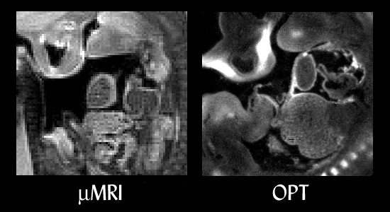 Close-up of the two reconstructions. A similar plane of section is shown from each embryo, at approximately the same magnification. Both embryos are facing left, with the front of the face at top left with nasal cavities visible in each, the branchial arch top centre, and tissues of the heart seen in the middle of each image. Thin membranes are not visible in the mMRI data. MRI data courtesy of R. Jacobs and S. Ruffins (see the
Caltech MRI mouse atlas project).
Close-up of the two reconstructions. A similar plane of section is shown from each embryo, at approximately the same magnification. Both embryos are facing left, with the front of the face at top left with nasal cavities visible in each, the branchial arch top centre, and tissues of the heart seen in the middle of each image. Thin membranes are not visible in the mMRI data. MRI data courtesy of R. Jacobs and S. Ruffins (see the
Caltech MRI mouse atlas project).